Alopecia Areata and /or Totalis and Vitiligo: An Expression Extraordinary of Multiple (Overlap) Autoimmune Syndrome
Juniper Publishers- JOJ Dermatology & Cosmetics
Letter to Editor

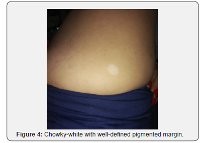
A 43-year-old mother of 3 young daughters was brought to the outpatients with the complained of excessive rarefaction of the scalp hair, following hair loss. The episode had its onset immediately after the delivery of the first child 15 years ago when her attention was drawn to a conspicuous, asymptomatic, hair loss localized at the temple of the scalp to which a little attention was paid. She was startled by the appearance of similar type of lesions in the adjoining part of the scalp coalescing to form a variegate configuration, ultimately progressing to involve practically almost whole (total) of the scalp, and embarrassing proposition, impelling her the use of wig as a camouflage, continuing its use until the next conception three and half year after. During pregnancy she had a substantive growth of hair span over a period of 9 month. The recurrence of events like that happening after first delivery concerned thepatients. The episode recitation too was a feature three and half year after third delivery and the same is being perpetuated up till now. Furthermore, she was in far yet another surprise, when she noticed the changing color (depigmentation/ whitening) on the lower part of the right thigh of 2 months duration. The patch was asymptomatic and progressive

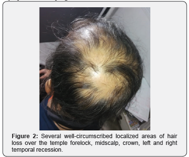
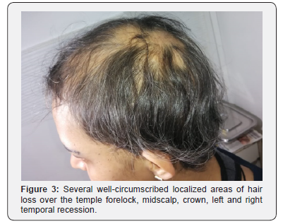
Examination of the scalp reveled well-circumscribed hair loss depicting rarefaction of the scalp. The hairs were thin and luster less. There were several lesions of identical morphology, coalescing to reflect variegated configuration. Baldness was apparent Figure 1-3. The lesions were present all over the scalp covering forelock, midscalp, crown, left and right temporal recession. In addition, chowky-white macule of the size of a rupee measuring 25mm was seen. Its borders were relatively pigmented and irregular. The lesion was located on posterior aspect of the right thigh above the ante-cubital fossa Figure 4. Endocrine functions test including thyroid; Tridothronine (T3)0.80ng/mL, Thyroxine (T4) 7.90ug/dL, Thyroid stimulating hormone(TSH) 3.16uIU/mL, parathyroid; Parathyroid hormone (PTH) 31.70pg/mL, adrenal cortex; adrenal function test adrenocorticotropic hormone (ACTH)<5.00pg/mL, pancreas; glycosylated hemoglobin (Hba1c) 5.6% and female gonads; folliclestimulating hormone (FSH)8.52mIU/mL , Luteinizing Hormone (LH)5.88mIU/mL, Prolactin (PRL) 8.90ng/mL test were undertaken and their values were within normal limits.


Multiple / overlap autoimmune syndrome is an intriguing overture, envisaging sound background in medicine in Journal and dermatology, warranting thorough workup of the patient. The initial description of syndrome was advance in the year 1988. [1] Describing simultaneous occurrence of three or more autoimmune diseases in the same patient indicating a pointer to a phenomenon predictive of autoimmunity. Based on critical evaluation of 87 cases available thus far brought into sharp focus a classification, the precise outlined of which are revived below in order to facilitate its applications in the future undertaking.
a) Type I comprises myasthenia, thymoma, polymyositis and giant cell myocarditis, this association having a single pathogenic mechanism.
b) Type II includes the Sjögren’s syndrome, rhumatoid arthritis, primary biliary cirrhosis, scleroderma and autoimmune thyroid disorders.
c) Type III groups together 10 autoimmune diseases (autoimmune thyroid disease, myasthenia and/or thymoma, Sjögren’s syndrome, pernicious anaemia, idiopathic thrombocytopaenic purpura, Addison’s disease, insulindependent diabetes, vitiligo, autoimmune haemolytic anaemia, systemic lupus erythematosus) for which a genetic predisposition seems to be an important factor
Undoubtedly, the classification is applied and practical. However, it is required to be modified to include one or more instead of three or more autoimmune diseases the contemporary accidences, bringing in its purview such cases thus enlarging its scope.
The current case is an illustration extraordinary embarrassing alopecia areata and /or totalis and vitiligo [2]. Alopecia areata/ totalis is one of the preeminent slowly progressive variants of alopecias, characterized by well circumscribed local loss of hair affecting primarily the scalp changing texture of the hair, thinning and rarefaction are its biological undertone, a major concern of the patient [3]. It is, therefore, imperative innovation to evaluate endocrine functions of thyroid, parathyroid, adrenal cortex, pancreas and female gonads to unfold their association with alopecia areata and /or vitiligo, and innovation required to be taken note in the future study of this nature.
Alopecia India is a well-established autoimmune [4,5] disease so also vitiligo [6]. Vitiligo and is association with other autoimmune diseases such as pemphigus vulgaris [7], with vitiligo [8] and myasthenia gravis has also been reported earlier. They too a retrospect (reclassified) into multiple/ overlap autoimmune syndrome.
The syndrome has been in reckoning ever since. However, its reporting has been too sporadic, seem to have gone into absolute oblivion for over a good decade until the year [9], focusing attention to yet another outstanding conglomeration vitiligo, alopecia areata, crohn’s disease, psoriasis vulgaris and oral lichen planus, having autoimmunity as a well-established pathogenesis [10].
For more Open Access Journals in Juniper Publishers please click on: https://juniperpublishers.com/
For more details JOJ Dermatology & Cosmetics (JOJDC) please click on: https://juniperpublishers.com/jojdc/index.php

Comments
Post a Comment