Neurophysiological Evaluation ff Paresthetic Notalgia- Juniper Publishers
Authored
by José Antonio Garbino
Abstract
Despite being a diagnosis little known to general
physicians, paresthetic notalgia is a frequent complaint in dermatology
clinic. The correlation with degenerative changes in the thoracic spine
is somewhat known, but the electrophysiological study in these cases is
still unfamiliar to the neurophysiologist. The objective of this paper
is to present a clinical case, describe and discuss the
neurophysiological evaluation of a patient with paresthetic notalgia.
Keywords: Diagnosis; physicians; paresthetic notalgia; Dermatology clinic; Hyperpigmentation
Introduction
Paresthetic Notalgia (PN) is a frequent reason for
searching a dermatology clinic. Due to the correlation of PN with
degenerative changes of the thoracic spine, neurophysiological
evaluation has been requested, but it is still an unusual entity for the
neurophysiologist. Paresthetic notalgia has been referred as: “dorsal
pruritus with hyperpigmentation”, “pruriginous dorsal melanosis”,
“Pierini and Borda melanocytic pruritus” [1], “localized hereditary
pruritus”, “pigmented posterior pruritic spot” and “pruritus
subscapular”[1,2]. For a long period it was considered as one of the
clinical varieties of primary cutaneous amyloidosis receiving the name
“dorsal amyloid lichen”[2]. It is now known that the presence of amyloid
substance is a consequence of pruritus and itching [1,3]. Paresthetic
Notalgia is considered as a chronic sensory neuropathy affecting in
particular the interscapular region between T2 to T6 levels, uni- or
bilateral, and may present a wider distribution [1,2].
Clinical Case
A 47-year-old female patient, who reported dark spots
on the upper dorsal region in both sides and on the lower right dorsal
region, accompanied by intense local pruritus, with a five-year
evolution. Background: chronic back pain and low back pain for 16 years.
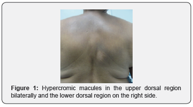
The dermatological examination showed hyperchromic
macules in the upper dorsal region bilaterally and the lower dorsal
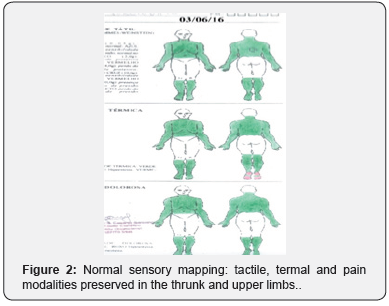
region on the right side, Figure 1. Normal nerve palpation; deep
reflexes normal and symmetric; sensory mapping: tactile, termal and pain
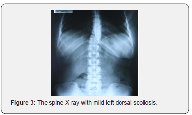
sensory modalities preserved, Figure 2. Image
examination: X-ray shows mild left dorsal scoliosis, without significant
kyphosis, Figure 3.
In neurophysiological approach the Electromyography
(EMG) was the technique chosen to evaluate the dorsal roots
because its has high sensitivity, specificity and reproducibility in
radiculopathies [4]. The EMG showed mild chronic neurogenic
motor pattern in dorsal paravertebral muscles of T3-T4 to T8-
T10 bilaterally and at the T8 and T9 level on the right side. In
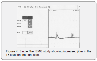
T5 level on the right, side increased jitter was recorded during
single fiber EMG study, characterizing a chronic denervation and
reinnervation process, with constant remodeling of the motor
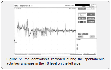
units [5], Figure 4. In T8 level, in the left side, pseudomyotonia
was found during spontaneous activities analyses, a feature of
axonal motor hyperexcitability, Figure 5.
Conclusion
In spite of being considered a sensory neuropathy [1,2], in
the this case of PN no sensory loss was found, tactile, thermal
and painful sensitivity in the involved territory associated with
Aβ, Aδ and C fibers was normal. However, the motor evaluation
showed root involvement in the symptomatic area. According to
the literature, pruritus would be mediated by C fibers, similar to
neuropathic pain, but due to the sustained hyperexcitability of
these fibers due to itching and to a lesser extent by posttraumatic
neuroplasticity [3]. These findings related to motor fibers
in dorsal roots would reflect a picture of global intraneural
hyperexcitability of the axons also involving nociceptive
C-related fibers related to pruritus, as postulated about A-wave
and pain in a previous papper [6].
For more
details JOJ Dermatology & Cosmetics
(JOJDC) please
click on: https://juniperpublishers.com/jojdc/classification.php
To read more…Full Text
in Juniper Publishers click on https://juniperpublishers.com/jojdc/JOJDC.MS.ID.555559.php

Comments
Post a Comment