Uncommon Elderly-Onset Nevus Lipomatosus Cutaneous Superficialis: A Case Report with Dermoscopic and Histopathologic Correlation-Juniper Publishers
Authored
by Suparuj Lueangarun


Abstract
Nevus lipomatosus cutaneous superficialis (NLCS) is a
rare hamartoma of the skin characterized by histological presence of
ectopic mature
adipocyte in the dermis. We presented an uncommon case of the
adult-onset classical form of NLCS, with the correlations between
dermoscopy
and histopathology of homogenous yellowish area, corresponding to the
histological finding of mature fat cell in upper dermisfor the diagnosis
of this type of skin condition.
Keywords: Nevus lipomatosus cutaneous superficialis; Hamartoma; Dermoscopy; Histopathology
Introduction
Nevus lipomatosus cutaneous superficialis (NLCS) is a rare
cutaneous hamartoma histologically characterized by the group
of mature adipocyte presenting among the collagen bundles of
the dermis. Clinically, this condition is classified into 2 types:
a) a classical or multiple type, usually presented at birth or
in the first two to three decades of life with the clinical group
of soft, painless, cerebriform, pedunculated, yellowish or
skin colored papules or nodules, and
b) a solitary type, usually developed in adults with solitary
papule or nodule. Most importantly, few cases of adult-onset
multiple types of NLCS were reported [1-3].
Case Report
A 74-year-old female presented with the 1-year history of
progressively increased numbers and sizes of asymptomatic
yellowish papules on the left upper thigh. She was otherwise in
good health even with her insignificant medical history. As well,
there was no history of local trauma, rubbing or scratching of the
affected sites, atopy or other significant underlying problems,
and no related Cutaneous problem in family members.Besides,
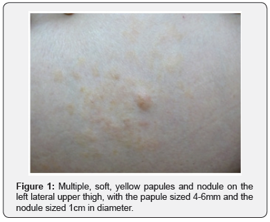
physical examination revealed groups of multiple, soft, smooth,
yellow-colored papules and nodule on left upper lateral thigh,
approximately 12 x 10 cm in size, and the largest nodule of 1cm
in diameter (Figure 1).

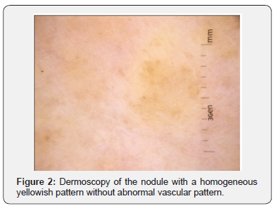
Whereas, the dermoscopic examination demonstrated
multiple well-circumscribed, homogeneous faint yellowish areas
without abnormal vascular structure (Figure 2). Moreover, the
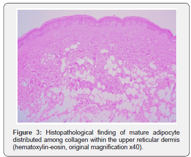
histopathologic examination from the largest nodule showed
multiple nests of lipocytes in the upper and mid dermis, with the
absence of skin appendages in this area, and normal overlying
epidermis (Figure 3). Hence, the diagnosis of nevus lipomatosus
cutaneous superficialis (NLCS) was confirmed.


Discussion
NLCS is a rare connective tissue hamartoma histologically
characterized by the presence of ectopic mature adipose tissue in
the superficial dermis. NLCS has been classified into two clinical
forms: a multiple or classic form and a solitary form [4]. The
classic form always presents at birth or develops during the first
two decades of life, clinically with grouped, fleshy, skin-colored
to yellowish papules and nodules on the lower trunk, buttocks or
thighs in a segmental distribution. Meanwhile, the solitary form
often occurs during adulthood as a solitary papule or nodule
anywhere on the skin. Interestingly, our case presented the
uncommon case of classical multiple form NLCS in the elderly,
[1-3] mostly found at birth or within the first two decade of life.
According to the postulated hypotheses of NLCS, degenerative
changes in dermal collagens and elastic tissues lead to fat cell
depositions among collagen bundles in superficial dermis [5]
and then precursor mesenchymal perivascular cells develop to
adipocyte in the dermis [6]. However, the genetic defect of NLCS
is still uncertain with only one cytogenetic reported study of the
2p24 deletion [7].
Our differential diagnoses include smooth muscle
hamartoma, plexiform neurofibroma, cutaneous leiomyoma,
and xanthogranuloma. Whilst, the definite diagnosis
requires histopathologic study to yield the characteristically
histopathologic finding of ectopic mature adipocytes lying
within the upper reticular dermis between collagen bundles and
around the blood vessels of the papillary dermis [8].
Additionally, the role of dermoscopyis crucial as a noninvasive
and useful tool for the diagnosis of many pigmented
and non-pigmented cutaneous lesions. In our present case, the
dermoscopic finding of homogeneous yellowish area corresponds
to the adipocyte in the dermis, and the lack of vascular structure
is also correlated with most components of mature fat cells in the
dermis with rare dilated capillaries from histology.
Furthermore, the yellowish color of fatty tissue findings from
dermoscopy also provide useful information for the diagnosis
of other skin conditions with adipocyte components such as
cutaneous lipomatous neuro fibroma [9]. However, the various
shades of homogeneous yellow color on dermoscopic finding
have been described in another condition including sebaceous
glands hyperplasia, tumors with sebaceous differentiation
[10], and dermal xanthomatous deposition diseases such
as xanthomatous dermatofibroma, reticulohistiocytoma,
and adult xanthogranuloma [11]. Particularly, Juvenile
xanthogranulomareveals the characteristically dermoscopic
findings of the ‘setting sun’ appearance with the orange-yellow
background of a subtle erythematous border [12].
In the meantime, treatment for NLCS is unnecessary, except
for cosmetic reasons. Following the conservative approach of
reported treatment, the lesions however tend to increase in
size and number, while occasionally present with rare ulcerated
complication [13].
Moreover, liquid nitrogen cryotherapy has been reported in
individual lesions with a partial response [14]. Nevertheless,
there is only one report that shows the effectiveness of CO2
laser ablation therapy for classic NLCS [15]. Thus, the treatment
of choice seems to be the surgical excision. However, the classic
form of clustered lesions is still limited, in term of large area
involvement.Also, no recurrence and malignant transformation
has never been before reported [4].
Conclusion
We report a rare case of adult-onset classical NLCS on
the left lateral upper thigh, with the dermoscopic finding of
homogenous yellowish area corresponding to the histological
finding of mature fat cells in the upper dermis for the diagnosis
implementation of this skin condition.
For more
details JOJ Dermatology & Cosmetics
(JOJDC) please
click on: https://juniperpublishers.com/jojdc/classification.php
To read more…Full Text
in Juniper Publishers click on https://juniperpublishers.com/jojdc/JOJDC.MS.ID.555552.php

Comments
Post a Comment